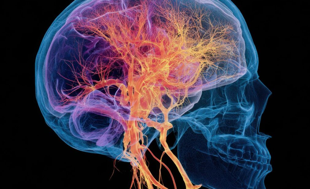
Vascular Imaging Department – Triplex Vascular & Arteriography
The Vascular Imaging Department of Ygeia Group offers specialized diagnostic services focused on the anatomical and functional assessment of the vascular system. Utilizing high-end ultrasound and angiographic imaging systems, the department ensures accurate diagnosis, early identification of vascular diseases, and optimal planning of vascular access, particularly in patients undergoing hemodialysis.
Core Services
- Vascular Ultrasound – Color Doppler Triplex
- Diagnostic Arteriography (Digital Subtraction Angiography – DSA)
- CT Angiography (CTA)
- MR Angiography (MRA)
- Preoperative Mapping for Arteriovenous Fistula (AVF)
- Surveillance of AVFs and vascular grafts
Color Doppler Ultrasound – Triplex
Color Doppler Triplex ultrasound is a non-invasive diagnostic method combining B-mode imaging, color Doppler, and spectral Doppler analysis to evaluate both vessel morphology and blood flow dynamics. It is essential for early diagnosis and monitoring of peripheral artery disease, venous insufficiency, carotid stenosis, and portal hypertension.
Applications:
- Carotid and vertebral arteries – stroke risk evaluation
- Peripheral arteries and veins – detection of stenosis, thrombosis, or reflux
- Cervical vessels – anatomical mapping and compression syndromes
- Splenoportal axis – assessment in portal hypertension and liver pathology
Arteriography for Arteriovenous Fistula (AVF)
Arteriography is a key diagnostic and interventional tool for patients undergoing or currently on hemodialysis. It is used for preoperative evaluation, fistula surveillance, and management of complications such as stenosis or thrombosis.
Indications:
- Preoperative vascular mapping for AVF or graft creation
- AVF maturation failure or dysfunction
- Thrombosis, low flow, or access-related complications
- Planning of alternative vascular access in complex cases
Imaging Techniques:
- 1. Digital Subtraction Angiography (DSA): High-resolution imaging with therapeutic capability (angioplasty, thrombolysis, stenting)
- 2. CT Angiography: Ideal for anatomical overview in complex cases
- 3. Duplex/Triplex Ultrasound: Real-time dynamic flow evaluation
- 4. MR Angiography: Preferred when contrast media or radiation must be avoided
Clinical Benefits
- Accurate and detailed vessel visualization
- Non-invasive or minimally invasive approach
- Potential for immediate therapeutic intervention
- Crucial for long-term planning of dialysis vascular access
- Multidisciplinary integration with vascular surgery, nephrology, and interventional radiology
Technology & Clinical Expertise
All examinations are performed by experienced vascular imaging specialists and interventional physicians using high-resolution ultrasound machines, angiography suites, and modern CT/MRI scanners. The department operates in close collaboration with Nephrology, Vascular Surgery, and Interventional Radiology departments.
Patient Instructions & Contact Information
- Fasting or renal function testing may be required for contrast-based or abdominal vascular exams.
- Most ultrasound exams require no preparation.
- Please contact the department for personalized instructions.