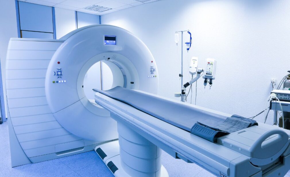
MRI Department
The MRI Department is engineered for high-level diagnostic and research needs, leveraging advanced hardware, optimized acquisition protocols, and continuous quality assurance. The technical architecture includes:
- Multi‑coil receiver arrays to enhance sensitivity and enable accelerated scanning (parallel imaging, SENSE/GRAPPA)
- Acceleration techniques (compressed sensing, undersampling) to accelerate scan times while preserving image fidelity
- Multipurpose multidimensional protocols (structural, diffusion, functional) as exemplified by recent implementations in population MRI studies
- Noise-suppression and motion-correction algorithms, crucial especially in pediatric and neuroimaging contexts
- Integration with PACS/RIS, compliance with DICOM/HL7 standards, and protocol standardization for interoperability
Acquisition protocols align with global guidelines (ACR, RSNA, ESGAR) and best practices in protocol harmonization .
Clinical Scope & Protocols
Applications span multiple domains:
1.
Neuroradiology
- T1, T2, FLAIR, SWI, DTI / tractography, and MR Spectroscopy for metabolic analysis
- Advanced susceptibility mapping (quantitative susceptibility mapping, QSM) to evaluate iron deposition in neurodegenerative disease
- Abbreviated/accelerated brain MRI protocols to reduce scan time in selected indications
2. Musculoskeletal MRI
- Cartilage, tendons, ligaments, bone marrow assessment via FS-PD, STIR sequences
- MR arthrography where indicated
- Protocols for osteomyelitis and soft tissue tumors
3. Cardiac MRI
- Ventricular function via cine SSFP
- Myocardial viability (Late Gadolinium Enhancement)
- Quantitative T1/T2 mapping
4.
Gynecological & Oncologic MRI
- Dynamic contrast-enhanced MRI for uterine/ovarian tumors
- Staging protocols (FIGO-compliant)
- Support for musculoskeletal and soft tissue oncology (DWI, perfusion)
5. Abdominal & Urogenital MRI
- MRCP for biliary and pancreatic imaging
- MR Enterography (MRE) for inflammatory bowel disease
- Quantitative liver imaging (PDFF, T2*) for steatosis/iron
- Dynamic and static uro-MRI for urinary tract evaluation
6. Pediatric MRI
- Age-optimized protocols for neuro, musculoskeletal, and abdominal imaging
- Motion-compensated and rapid-acquisition techniques
Quality Assurance, Safety & Innovation
- Implementation of formal quality control protocols (technical QC, radiological QC)
- Continuous monitoring of safety issues (thermal burns, acoustic noise) and staff training
- Management of risks associated with static, RF, and time-varying gradient fields
- Adoption of novel methods (deep learning) for accelerated reconstruction and motion correction
Examination Workflow & Clinical Support
- Pre-scan triage to define clinical indication and protocol alignment
- Engagement with patient for pre-scan instructions (e.g. implants, contrast history)
- Selective contrast administration according to protocol
- Real-time image transfer and evaluation on PACS
- Structured reporting and liaison with the referring physician
Contact the nearest center for referral submission and scheduling
Bibliography
- Eisenmenger LB et al. Focused abbreviated survey MRI protocols for brain and … Radiology / RSNA (2023).
- Sharma PS et al. Standardizing Magnetic Resonance Imaging Protocols: addressing challenges in diagnostic accuracy. Journal of Magnetic Resonance Imaging (2020).
- Shetty AS et al. Body MRI: Imaging Protocols, Techniques, and Lessons. Radiographics / RSNA (2022).
- A narrative review of current and emerging MRI safety issues. PMC (2023).
- Deep Learning for Accelerated and Robust MRI Reconstruction: a Review. (Heckel et al., 2024)
- Deep Learning for Retrospective Motion Correction in MRI: A Comprehensive Review. (Spieker et al., 2023)
- A Comprehensive Literature Review of Application of fMRI in Healthcare. (2021)
- Design and validation of the ADNI MR protocol. (Arani et al., 2024)
- Design and validation of the ADNI MR protocol. (Arani et al., 2024)