Specialized Digital Radiology Department
Full-Spine Digital Radiography
Full-spine digital radiography is a standard imaging technique for evaluating spinal deformities such as:
- Idiopathic or secondary scoliosis
- Kyphosis (juvenile, degenerative, or post-traumatic)
- Lordosis and other spinal axis deviations
Performed in a single, full-length projection, the scan captures the entire spine from the skull base to the pelvis.
We use a high-resolution, low-dose digital system, ensuring excellent image quality with minimal radiation exposure in accordance with ALARA principles.
Full-Length Lower Limb Radiography
This imaging modality is widely indicated for:
- Evaluation of limb length discrepancy
- Assessment of lower limb mechanical axis
- Pre- and post-operative control after orthopedic procedures (e.g. hip or knee arthroplasty)
It enables accurate anatomical and mechanical axis measurements, aiding in surgical planning and follow-up.
No patient preparation required.
Contact us for appointment availability and pricing.
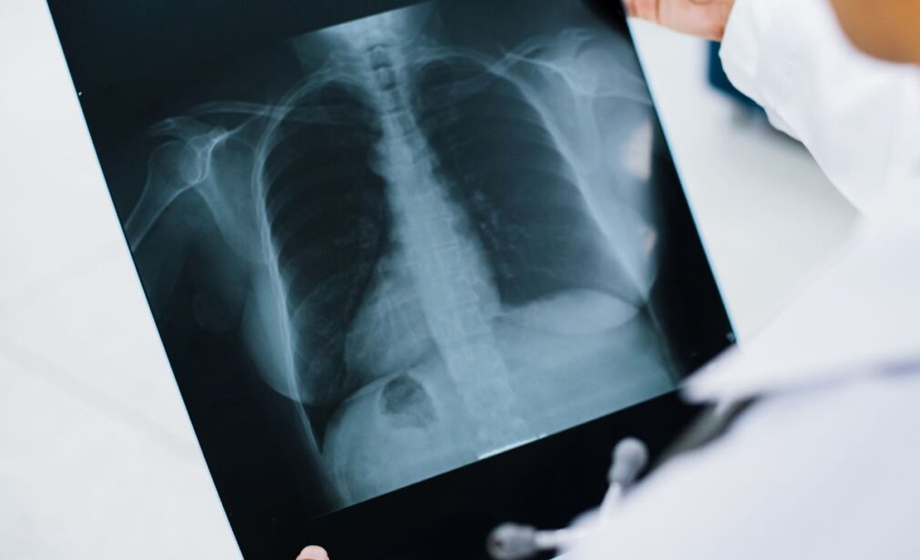
General Digital X-rays
Routine digital radiographs are performed daily in all essential anatomical areas:
- Chest
- Paranasal Sinuses
- Spine (Cervical – Thoracic – Lumbar)
- Bones & Joints
- Kidneys – Urinary Tract (KUB)
- Skull
- Pelvis – Sacroiliac Joints – Hips
- Abdomen
Digital radiography provides:
- High-resolution images
- Significantly reduced radiation dose
- Immediate electronic access to results
Panoramic & Cephalometric Radiography
Digital Orthopantomograph – Dental & Maxillofacial Imaging
Our Radiology Department is equipped with a modern digital orthopantomograph, ideal for:
- Panoramic Radiographs (OPG): Full dentition assessment, impacted teeth, cysts, jaw fractures.
- Cephalometric Imaging: Essential for orthodontic planning and craniofacial structural analysis.
Low-dose imaging protocols make this technology safe for children and adolescents, with results available digitally.